Knee Pain in Sweden: 3 Guided Self-Tests & When to Consider Private MRI
Clinically reviewed guide · Updated
If you develop knee pain in Sweden, you may be unsure where to seek care. The public primary care healthcare queue (vårdcentral) can take several weeks for an initial appointment, and obtaining an MRI referral often involves months of waiting. Yet early differential diagnosis is critical to preventing long-term cartilage or ligament deterioration.
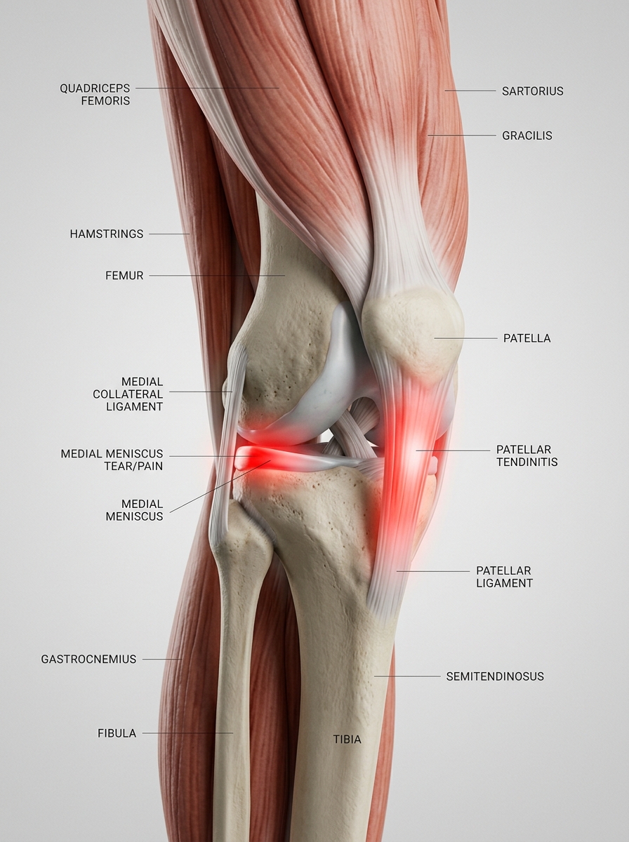
Mapping Your Symptoms: Three Common Presentations
Before undergoing formal clinical examination, pinpointing the mechanical location provides strong diagnostic cues:
- Anterior (behind or below the patella): Characteristic of patellofemoral pain syndrome (runner's knee) or patellar tendinitis. Aggravated by descent on stairs or prolonged sitting.
- Medial or lateral joint line: Highly indicative of meniscal pathology or collateral ligament strain. Usually provoked by rotational twisting.
- Generalized aching with morning stiffness: Typical of early joint cartilage wear (knee osteoarthritis).
The 3 Clinician-Designed Self-Tests
Perform these functional tests gently on a flat, even surface. Cease immediately if you experience sharp, acute pain.
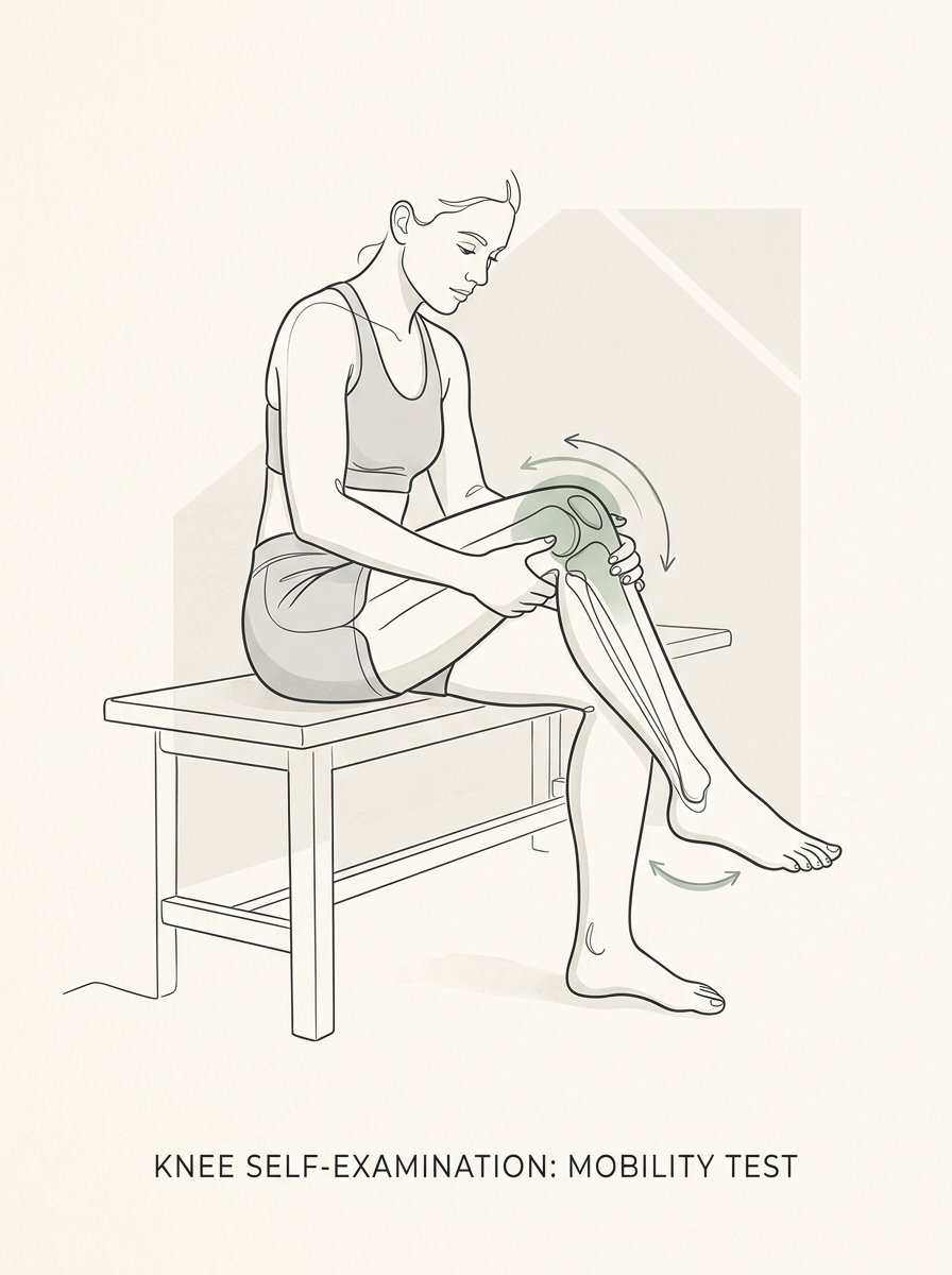
Test 1: Step-Down Descent Test
Execution: Stand on a sturdy 15–20 cm step. Slowly reach forward with your unaffected leg toward the floor, bending the affected knee under controlled single-leg loading.
• Localized anterior aching behind the kneecap during descent points to patellofemoral tracking stress.
• Deep catching or sharp pinching along the inner margin suggests meniscal compression.
Test 2: Gentle Torsional Load Test (Thessaly Variation)
Execution: Stand barefoot with light finger support on a wall or desk. Bear approximately 70% of your body weight on the affected leg with the knee slightly bent (20° flexion). Slowly rotate your hips and torso inward and outward twice.
• Joint-line pinching, clicking, or an inability to complete the rotation strongly correlates with meniscal involvement.
Test 3: Morning Stiffness Index
Execution: Monitor joint behavior during the initial 30 minutes after waking or following prolonged desk work.
• A stiff sensation that gradually clears with 10–15 minutes of light movement, followed by evening aching, is a classic presentation of early osteoarthritis.
Differential Comparison Table
| Condition | Primary Pain Site | Aggravating Factor | Recommended Next Step |
|---|---|---|---|
| Meniscus Tear | Medial or lateral joint line | Twisting, deep squats, pivot sports | Physical therapy + MRI if locking occurs |
| Osteoarthritis | Diffuse, frequently medial | First morning steps, cold weather | Structured strength protocol, weight-bearing X-ray / MRI |
| Runner's Knee (PFSS) | Behind/under kneecap | Downhill walking, stairs, running | Targeted hip/quadriceps kinetic rehab |
Vardse Nordic: Direct Video Assessment & Private MRI in Sweden
If your self-tests indicate structural meniscal or ligament pathology, you do not need to wait through public queues.
- Digital Physiotherapy Evaluation (60 min): Comprehensive video examination with an English-fluent licensed Swedish physical therapist within 24–48 hours.
- Physician Review: Medical assessment and referral for advanced diagnostic imaging.
- Private Knee MRI: Arranged through partner imaging centres in Stockholm, Gothenburg, and Malmö with appointments within 10 business days.
- Personalized Recovery Plan: Full follow-up, specialist imaging review, and surgical referral if necessary.